Health Scan: Better pediatric simulation system in Tel Aviv

Individuals with severe overweight issues have an inhibited sense of satiation; they release fewer satiety hormones than people of normal weight.
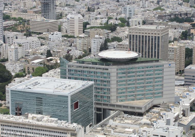
Ichilov hospital and Sourasky Medical Centre in Tel Aviv. (photo credit: WIKIMEDIA COMMONS/GELLERJ)
Ichilov hospital and Sourasky Medical Centre in Tel Aviv.
(photo credit: WIKIMEDIA COMMONS/GELLERJ)
A simulation system for children that uses up to 20 times less radiation than computerized tomography (CT) and at a speed of only 10 seconds has been introduced at the Dana-Dwek Children’s Hospital at Tel Aviv Sourasky Medical Center. The EOS imaging system, based on a invention that won the Nobel Prize in Physics for the low dose of particles, has been introduced here so far only at the children’s hospital.
The EOS system is groundbreaking in that it scans the patient while standing and enables a 3D reconstruction of the photographs. The system absorbs x-ray scanning performed simultaneously on two levels and then is able to integrate the two planes and perform the same three-dimensional reconstruction using ultra low-dose radiation.
The EOS system emits up to two to three times less radiation than general radiography and 20 times less than computed tomography (CT) and in backlit images, up to 85% less radiation than in computerized radiography of equal or better quality. In lower extremities and spinal column length, the radiation dose is reduced by 50% compared to computerized radiography; in axis and lower-limb structure lesions, it requires a radiation dose 95% lower than CT. There is also a protocol for performing a low-dose dose test in which all the doses listed above are reduced 10-fold.
Dr. Dror Ovadia, director of the pediatric orthopedics department, says, “Reducing the radiation dose is especially beneficial for children who need to take photographs frequently, such as children with spina bifida or scoliosis. For us, the innovative system is the best option to monitor children and monitor disease progression.”
The device also reduces the time it takes to perform the scan to only 10 to 25 seconds. EOS technology provides real-size, 1:1 scale images for surgical design and monitoring of bone and joint diseases.
“The three-dimensional images of standing children enable us to accurately see the spine and lower limbs, the interaction between the joints and the rest of the skeletal system, in particular the spine, hips and legs, so we can assess the balance and stability and examine the bones, joints and ligaments from several angles,” he explained.
“This kind of simulation enables us to see all the regions of the body in one photograph instead of combining several pictures together. Such imaging allows accurate visualization of the musculoskeletal system necessary for diagnosis and treatment planning.”
The EOS device is primarily used to evaluate patients’ spinal, hip, knee and lower extremities. One of the well-known limitations of studies performed on spinal deformities is the use of 2D imaging. The idiopathic scoliosis (whose cause is not known) is called a threedimensional deformity of the spine. Routine scoliosis does not allow accurate evaluation of the axial dimension (the rotation of the vertebrae) and often underestimates the deformity created by the vertebrae’s structure. The EOS system, which allows simultaneous shooting of two planes and performing 3D reconstruction later, results in improved measurements and a better understanding.
OBESE PEOPLE LACK SATIETY HORMONES
Individuals with severe overweight issues have an inhibited sense of satiation; they release fewer satiety hormones than people of normal weight. The reason is that there are fewer cells producing satiety hormones in the gastrointestinal tract of the obese, according to Swiss doctors writing in the journal Scientific Reports. Surgical weight-loss procedures can repair this disorder.
The mucous membrane of the upper gastrointestinal tract is home to highly specified cells, the so-called enteroendocrine cells, which constantly analyze our intestinal contents. During a meal, they release satiety hormones into the bloodstream, signaling to the body that enough food has been taken in and that the meal can be ended. The sense of satiation is created in the central nervous system. Compared to lean people, the release of satiety hormones is reduced in obese people. After bariatric surgeries such as a gastric bypass or sleeve gastrectomy, the release of these hormones increases again.
Researchers from the biomedicine department at the University Hospital of Basel and the St. Claraspital private hospital in Basel, together with colleagues from the University of Liverpool, have studied the reasons for this reduced release of satiety hormones. They examined tissue samples of the gastrointestinal tract in 24 lean volunteers and 30 obese patients before and after weight-loss surgery.
The team of researchers, led by Dr. Bettina Wölnerhanssen, was able to show that the number of enteroendocrine cells in obese people is significantly lower than in people with normal weight. This leads to a reduced release of satiety hormone which in turn leads to altered appetite. The obese also showed alterations in the pattern of the so-called transcription factors that are responsible for the development of enteroendocrine cells from stem cells. After surgery, the number of enteroendocrine cells and the pattern of transcription factors were almost entirely restored.
“Unfortunately, obese people are often stigmatized, and it is a common belief that the reason for obesity lies in a lack of self-control and discipline,” commented Wölnerhanssern. However, there is no doubt that metabolic factors are playing an important part. The study shows that there are structural differences between lean and obese people, which can explain lack of satiation in the obese.