
Researchers from the Weizmann Institute of Science discovered a new method for mapping gene expression deep within the body, and their findings may completely change the way we approach observing the biological processes of the human body in the future.
Our cells have many biological functions that influence gene expression, though many of these functions have yet to be properly observed, as scientific methods for viewing them are currently lacking. Scientists typically use colorful glowing proteins to create contrast within cells so that they can better see the intricacies inside of them, but these proteins are insufficient when it comes viewing to processes that occur more deeply within the body.
That may be changing soon, however, thanks to a new peer-reviewed study by Dr. Hyla Allouche-Arnon and a team of researchers from the Bar-Shir lab at the Weizmann Institute of Science. Together they demonstrated that it is possible to observe these deep cell processes with the help of magnetic resonance imaging (MRI). The bodily tissues that obscure cell functions when glowing proteins are used for contrast do not impede the radio-wave signals that MRIs emit.
In their study, the researchers developed a method for using MRI technology to track the expression of two genes using different colors, a groundbreaking technique that lays the groundwork for future use of MRI as a tool for examining other biological processes non-invasively, including "monitoring the effects of cancer therapy or tracing stem cells introduced into the body for therapeutic purposes," according to a press release from the Institute.
Dr. Amnon Bar-Shir of the Molecular Chemistry and Materials Science Department and head of the research team explained: "MRI may one day be used to peer deep into the body over an extended period of time, to see what goes on in tissues without the need to remove them for study under a microscope. Our method provides a major step in that direction."
Ahead of his time, Nobel laureate Roger Y. Tsien actually predicted that MRI could one day take the place of glowing proteins for viewing gene expression during his 2008 acceptance speech, though at the time MRI was not capable of detecting several genes at once through differentiative colors.
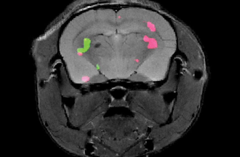
This is where the team from Weizmann came in. They developed a method for detecting multiple genes by genetically engineering two groups of cells that would each express a unique, specially designed protein. Simultaneously they combined two different molecular probes that would later be injected into the bloodstream of mice so that they would accumulate only in the cells that expressed the engineered proteins. The probes were designed to light up in pink or green in response to particular frequencies from an MRI scan, which enabled the team to detect the exact positions of the engineered cells.
Allouche-Arnon explained what this means for the future of gene expression mapping, “Gene expression lets us know what each cell is doing. Thanks to our method, MRI may now be applied by researchers in various fields to track the activity of all kinds of processes, for example, those involving different types of brain or immune cells.”
Study participants also included Nishanth D. Tirukoti, a research student in Bar-Shir’s lab; Dr. Yoav Peleg, Dr. Orly Dym, Dr. Shira Albeck, Dr. Alexander Brandis and Tevie Mehlman of Weizmann’s Life Sciences Core Facilities Department; Dr. Liat Avram and Dr. Talia Harris of Weizmann’s Chemical Research Support Department; and Dr. Nirbhay N. Yadav of the Johns Hopkins School of Medicine.
Dr. Amnon Bar-Shir is the incumbent of the Helen and Milton A. Kimmelman Career Development Chair.
Dr. Bar-Shir’s research is supported by the Clore Institute for High-Field Magnetic Resonance Imaging and Spectroscopy; and the Helen and Martin Kimmel Institute for Magnetic Resonance Research.