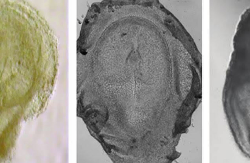
No one would ever confuse a human being with a mouse, a chicken, or an elephant, but at the very start of their different developmental paths, they share a striking similarity. In fact, even the most experienced researchers have a hard time saying whether an embryo in its gastrulation stage – one of the earliest stages of embryonic development – will turn out to be a creature that talks, squeaks, cackles, or trumpets.
The three germ layers during gastrulation are the endoderm, the ectoderm, and the mesoderm. Cells in each germ layer differentiate into tissues and embryonic organs. The ectoderm gives rise to the nervous system and the epidermis, among other tissues. The mesoderm gives rise to the muscle cells and connective tissue in the body. The different species’ embryos begin their lives from different starting points and, of course, develop into species with their own unique organs, characteristics, and sizes.
But at one particular moment, they surprisingly display remarkably similar shapes. Biologists describe this phenomenon using the hourglass model: Like the sand that flows from bulb to bulb, the development of embryos of every species of vertebrates – including mammals, fish, birds, and reptiles – passes through a narrow bottleneck, at which point they are all almost identical.
Since it is so problematic to investigate human embryos at the earliest stage of development, scientists make use of animal models, mostly rodents. But after almost a century of extensive research into mouse embryos, which has produced many important discoveries, we still know very little about the very beginning of human life – and it is far from certain that the embryonic development pattern of mice is similar enough to the same process in humans.

A study by researchers at the Weizmann Institute of Science in Rehovot was recently published in Cell under the title “Time-aligned hourglass gastrulation models in rabbit and mouse” that proposed a new approach to understanding the mystery of how embryos are created. The study – by Dr. Yonatan Stelzer and Dr. Yoav Mayshar of the molecular cell biology department and Ofir Raz and Prof. Amos Tanay of the computer science and applied mathematics and the molecular cell biology departments – uncovers new information about the earliest stages of embryonic development and could help answer additional key questions about the matter.
Showing how an embryo is formed
The researchers used a system they developed in an earlier study conducted on mice that, for the first time, successfully described the process of embryonic development over time. The system relies on information collected from tens of thousands of individual cells with pictures and physical measurements of individual embryos. The researchers managed to take many such snapshots to create a kind of movie that shows – hour after hour – how an embryo is formed.
In the current study, they used the same system to address a significant challenge, one that scientists had been forced to overlook until now: the fact that embryonic development in mice differs from that of most other mammals, including humans. While mouse embryos form an elongated cylinder during the gastrulation stage, other mammalian embryos resemble an almost flat disk. This fundamental geometric difference decides the different locations of the embryonic cells and tissues at the critical moments when the cells differentiate into future nerve cells, reproductive cells, or those that will form the digestive system.
The differences and similarities among mouse embryos and those of other mammals raise fascinating questions that, until now, were difficult to address: How are the cylindrical mouse embryos and the disk-like embryos alike in terms of gene expression and cellular development, and how do they differ? How do genes and cells of such different creatures converge and display similar behavior at a certain developmental moment? And is there any species that could serve better than mice as a model to crack the secrets of human development?
To find answers, the researchers went down the rabbit hole. Although rabbits are very different from mice, they do share some characteristics that make them valuable animal models. At first, the researchers repeated the process that they had performed on mice, mapping the gene expression and the development of cells and tissues as they changed over time. They then began identifying the precise characteristics of each cell and every developing tissue.
“We used computer models to identify genes and to characterize the types of cells activated in the first stages of the rabbit’s embryonic development,” Tanay explained. “Cutting-edge technological tools allowed us to reach, in a relatively short time, the highest levels of detail and accuracy, which had taken decades to obtain in studies of the embryonic development of mice.”
Once that complex mission was completed, the researchers had two “movies” that enabled them to compare the real-time embryonic development of the two different species. This extremely significant comparison produced illuminating findings. First, the scientists found striking similarities between mice and rabbits in the expression of genes responsible for tissue development at the convergence stage described by the hourglass model – and they successfully identified around 75 genes that are key factors in this process. This discovery validates the hourglass model and shows that, despite the significantly different geometrical shapes of the embryos, their genes and cells behave in a remarkably similar way. That, in turn, provides researchers with a first clue regarding the evolutionary processes that, during the gastrulation stage, preserve a similar embryonic shape in different species, from which a huge variety of shapes later develops.
The researchers also identified massive differences between mice and rabbits when it came to the development of early reproductive cells, later to become the egg and sperm – differences that are of key importance to nature and scientists alike because they are essential for maintaining continuity across generations. A deeper understanding of how these cells develop in rabbits could provide a better understanding of the same process in humans. Another major difference between mice and rabbits was found in the extraembryonic tissues, which are vital for the proper development of the embryo – the placenta and the yolk sac.
“This study presents computational and theoretical tools that provide fertile ground for future research,” Stelzer concluded. “The ability to compare species at corresponding points in time in embryonic development is hugely important for understanding evolutionary processes. Moreover, the new information about rabbits, which is apparently more relevant to humans than the data gleaned from mice over the years, can shed light on the earliest stages of human embryonic development and contribute to applied and medical research.”
