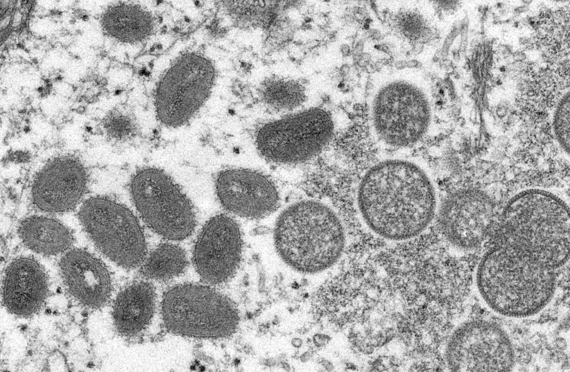
A 31-year-old man diagnosed with Monkeypox developed acute myocarditis, implicating heart inflammation as a possible complication of the viral disease, according to a new study published in JACC: Case Reports.
Monkeypox is caused by the monkeypox virus, which is related to the virus that causes smallpox and is transmitted through close contact with bodily fluids, respiratory droplets or lesions. Like smallpox, symptoms of monkeypox may include pimples or blister-like rashes on the hands, feet, face or genitals.
The patient went to a health clinic five days after developing symptoms of the disease, including fever, lesions, myalgia and malaise, and was diagnosed with Monkeypox using a PCR swab of a lesion. Three days later, the patient returned to the clinic reporting chest tightness that could be felt in the left arm.
According to the study, a combination of clinical presentation and noninvasive diagnostic findings such as cardiac magnetic resonance (CMR) abnormalities can be used to diagnose myocarditis.
An ECG showed cardiac abnormalities and lab tests showed increased levels of C-reactive protein, high-sensitivity troponin I, creatine phosphokinase (CPK) and brain natriuretic peptide (BNP), potential markers of heart injury.

“Through this important case study, we are developing a deeper understanding of monkeypox, viral myocarditis and how to accurately diagnose and manage this disease,”
Julia Grapsa, MD, PhD, editor-in-chief of JACC: Case Reports
Julia Grapsa, MD, PhD, editor-in-chief of JACC: Case Reports said that researchers used CMR mapping, a method of imaging that the researchers noted is "the noninvasive gold standard for the diagnosis," to confirm that the patient had myocardial inflammation and acute myocarditis.
“Through this important case study, we are developing a deeper understanding of monkeypox, viral myocarditis and how to accurately diagnose and manage this disease,” she said.
Status of the patient
The patient was discharged from the clinic after making a full recovery in one week. The researchers noted that more research is needed regarding the relationship between monkeypox and heart injury.
"We believe that reporting this potential causal relationship can raise more awareness of the scientific community and health professionals for acute myocarditis as a possible complication associated with Monkeypox and might be helpful for close monitoring of affected patients for further recognition of other complications in the future," the study read.
