Alzheimer’s disease – the progressive neurological disorder that causes the brain to shrink and brain cells to die – is the most common cause of dementia. The disease causes a continuous decline in thinking, behavior and social skills that affect a person’s ability to function independently.
But while the disorder is incurable, it is important to diagnose it as rapidly as possible so measures can be taken to slow the decline. Doctors hope to eventually develop treatments to reduce the risk of developing Alzheimer’s disease.
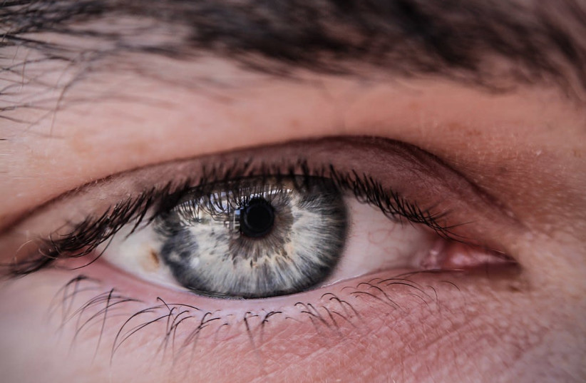
But now, doctors in the ophthalmology department of the Samson Assuta-Ashdod University Hospital suggest a much simpler way to diagnose Alzheimer’s – by looking for beta-amyloid plaques and abnormal tau proteins in the retina of the eye. The advantage is the accessibility of the retina for direct visualization by non-invasive means.
The research, just published in the latest issue of Harefuah – the Hebrew-language journal of the Israel Medical Association – was conducted by Drs. Keren Wood of the Samson Assuta Ashdod Hospital and Ben-Gurion University of the Negev, Idit Maharshak of Wolfson Medical Center in Holon and Tel Aviv University’s Sackler Faculty of Medicine, and Yosef Koronyo and Maya Koranyo-Hamaoui of the Cedars-Sinai Medical Center in Los Angeles, California.

How can you diagnose Alzheimer’s through the retina?
The retina is a component of the central nervous system that can easily be accessed by technology used routinely by ophthalmologists, they wrote. Photoreceptors in this “screen” at the back of the eye absorb light and transfer data to the retinal ganglion cell layer. Axons (long, slender nerve fibers) in this layer accumulate along the retinal nerve fiber layer and transfer the data to the brain via the optic nerve connected to the eye.
Since the retina is connected to the brain, it seems that changes in this part of the eye reflect pathological processes in the brain, the authors wrote, including the development of Alzheimer’s disease. Amyloid-beta plaques have been found in the retina of cadavers in autopsies of people who died of Alzheimer’s.
Turmeric is a natural, intensely yellow-colored spice that attaches itself to plaques of amyloid-beta. Ten Alzheimer’s patients and six healthy controls were asked to swallow turmeric capsules. A few days later, their retinas were examined. The yellow spice was found to stick to the retinal cells in Alzheimer’s patients but not in the healthy controls.
Other non-invasive tests of the retina – including optical coherence tomography and optical coherence tomography angiography – were also conducted and found to point to the early development of Alzheimer’s, the authors wrote. Still, larger tests must be conducted with these means before they can be implemented clinically. A clear biomarker must also be found in the individual to be sure the patient is developing Alzheimer’s and sent for treatments, they concluded.