
Tissue engineering, 3D printing and a lot of hope were the keys for researchers at the Technion - Israel Institute of Science and Sheba Medical Center to engineer an ear.
What does it take to make an ear? Researchers developed an efficient technology to allow the production of functional, customized aesthetic implants for the rehabilitation of improperly developed ears.
Microtia is usually restored using rib cartilage tissue taken from the patient's chest area. This method involves pain and discomfort, as well as the risk of further complications. Moreover, the creation of an ear that is exactly similar to the other ear is dependent on the surgeon's creative skills and advanced surgical abilities, and therefore isn’t always possible. In the current study they tried to find a new, effective and less painful method.
The researchers, whose work was published in the journal Biofabrication, were able to meet this goal by incorporating new tissue engineering technologies, developed at the Levenberg Laboratory led by postdoctoral fellow Dr. Shira Landau, where they developed a biodegradable skeleton which grew an ear.
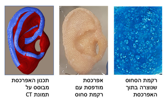
The unique skeleton, which allows the creation of an aesthetic and stable outer ear, is produced in three-dimensional printing according to the CT image. It’s a biodegradable skeleton on which chondrocytes, which are specialized cells from which cartilage is made, grow along with microchemical stem cells. The skeleton contains tiny perforations of various sizes that allow the cells to attach to each other and form stable cartilage tissue.
The researchers monitored the growth of cartilage in the skeleton in the lab for 10 days to six weeks and then implanted the ear in a mouse model. The result was a successful implant absorption and good biomechanical function of the artificial ear.
Researchers explained that creating an engineered earpiece (auricle receiver) from the patient's own cells would reduce the suffering and risk caused to children as a result of harvesting rib cartilage from their bodies. In addition, it will allow the surgeries to be performed as early as around age six and not after age 10, as is customary today. Surgery at a younger age may reduce the psychological consequences of microtia in children, since they can start first grade looking like the other kids.
This breakthrough was achieved in collaboration between Levenberg; Dr. Shay Izhak Duvdevani, a senior physician in the Department of Otolaryngology and Head and Neck Surgery and Head of the Tissue Engineering Laboratory at Sheba; and Dr. Orit Harari Steinberg, director of the Tissue Engineering Laboratory at Sheba.
Levenger explained that one challenge in the study was to find a suitable method for 3D printing, since the creation of an ear requires the use of materials that will be absorbed in the body. So, there’s a very precise external structure with tiny holes. She added that all these results were shown in the present study, and they believe that it will be possible to adapt this technology to other applications such as nose rehabilitation and the creation of various orthopedic implants.
Duvdevani noted that “in this study we have reached a significant breakthrough thanks to the integration of the clinical world into the research world and the collaboration between physicians and researchers.
“This study is another milestone in the transition to advanced technologies in medicine, in which the use of 3D and tissue engineering will play a significant part and will provide an optimal and advanced solution for patients.”