 communicate with vessels located in the surrounding hydrogel (green).")
Skin flaps, bone grafts, implanted tissue - recent advancements in medicine have changed the face of surgery in terms of autologous - meaning self - transplantations.
While extensive damage to organs once meant a nearly sure amputation or need for an external transplant, today's science focuses on harvesting cells and tissue from a person's own body to complete the injured pieces of the puzzle, using grafts and flaps to repair skin, vessels, tubes and bones.
Yet, ask any surgeon attempting to insert a flap and they would tell you that the most important - and restrictive - component of a graft's success is ample blood supply.
A team of researchers at the Technion recently found a way to meet this need. For the first time, these scientists succeeded in 3D printing a network of big and small blood vessels that could provide blood to implanted tissues just like the human body.
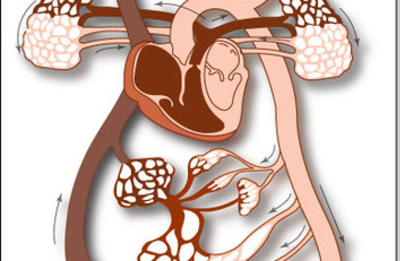
Up until now, medicine hasn't been able to mimic the body's ability to create a suitable hierarchy in the blood vessel tree. In our bodies, the heart pumps blood into a large tube called the aorta, which measures roughly 2-3 cm in diameter. The blood vessels then branch off into smaller and smaller tubes that are appropriate to each organ's need and capacity, until they reach minuscule arterioles of only 5 to 10 micrometers.
Dr. Ariel Alejandro Szklanny of the Technion team, led by Professor Shulamit Levenberg, a specialist in tissue engineering, found a way to use 3D printing to form a system containing a functional combination of both the large and small vessels.
The new breakthrough may allow a tissue flap to be created in a lab already connected to a blood network suited to its size and function.
Currently, transplanted grafts need to be implanted into a healthy part of the body so that the patient can generate new blood vessels to support it; then, the graft is relocated to an affected area as healthy tissue.
The new technique could potentially eradicate this intermediate step, drastically improving recovery times and cutting down on the number of procedures a patient would need to undergo.
In his recently published study in Advanced Materials, Dr. Szklanny described how he created a polymeric scaffold filled with small holes, mimicking the large blood vessels of the body. These holes allowed the connection of smaller vessels to join into the engineered large vessels. With collagen bio-ink, the team then printed and assembled a complex network around and within the main scaffold, later covering it with endothelial (human blood vessel lining) cells. A week later, the incubated artificial apparatus joined with the cells to create a hierarchical structure just like the human blood vessel tree.
While previous studies in this field used animal-borne collagen, the Technion team used engineered tobacco plants created by the Israeli company CollPlant.
The mesh was transplanted into a study rat and attached to the main artery in its leg. The blood through the artery spread through the network exactly as it would within the body, carrying oxygen and nutrients to the distant parts of the implanted tissue, and without any leaks.
This achievement is an important tool in the world of personalized medicine and could be a huge leap forward in tissue engineering and treatment.