
Embryonic stem cells (ESCs) have great potential in the field of regenerative medicine and tissue engineering because they have the ability to produce every type of cell and tissue in the body.
Researchers at the Hebrew University of Jerusalem (HU) and the University of Toronto have harnessed fluorescent proteins (FPs) to investigate changes in the cell nucleus during embryonic stem cell differentiation.
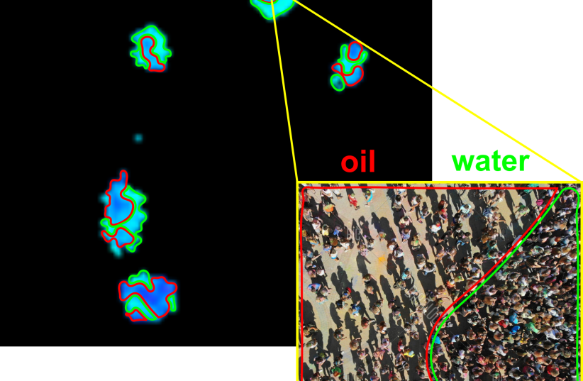
Their discovery provides important information about how our cells work and change over time, the scientists said. Specific features of FPs called fluorescence lifetimes acted as beacons revealing how biomaterials are packed within the nucleus of cells.
They found that as embryonic stem cells mature, these biomaterials become more uniformly distributed, resembling oil droplets in water, but with intriguing complexities.
Fluorescent proteins as a scientific game-changer
Fluorescent proteins have long been a staple in cellular research, opening for scientists a glimpse into the inner world of cells, but this study introduces an innovative application of FPs, allowing them to serve as indicators of local density within bio-condensates in the cell nucleus.
This innovative method opens new avenues for understanding cellular development – the body’s raw materials – as under the right conditions in the body or a lab, stem cells divide to form more cells called daughter cells, the team said.
ESCs are found in the inner cell mass of the human blastocyst – an early stage of the developing embryo that lasts from the fourth to the seventh day after fertilization. In normal embryonic development, they disappear after the seventh day and begin to form the three embryonic tissue layers.
The new study, just published in the prestigious peer-reviewed journal Nature Communications under the title “Fluorescent protein lifetimes report densities and phases of nuclear condensates during embryonic stem-cell differentiation,” reveals a powerful new method for studying the inner workings of cell nuclei during embryonic stem cell differentiation.
The team, led by Dr. Eitan Lerner from HU’s Institute of Life Sciences and the Center for Nanoscience and Nanotechnology and Prof. Eran Meshorer, from that institute and the HU’s Edmond and Lily Safra Center for Brain Sciences (ELSC and Prof. Sarah Rauscher from the University of Toronto used a specific feature of special glowing proteins to learn about how cells change and grow.
These protein-based “reporters” helped them understand how parts of DNA called heterochromatin are packed inside the cell nucleus and what happens to these parts during cell development.
Commenting on their study, Meshorer said “Our research opens new doors for understanding the complexities of cellular behavior during differentiation. The ability to precisely track local densities within bio-condensates using fluorescent proteins provides valuable insights into cellular development that were previously hidden from view.”
Lerner added that “this breakthrough method offers researchers a powerful tool for investigating the intricate processes that underlie critical cellular events in general and stem cell differentiation in particular. This discovery paves the way for gaining a better understanding of the inner cell intricacies, and, as such, has the potential to reshape our understanding of cell biology.
It represents a significant step forward in the field of cellular biology and holds promise for future applications in understanding various cellular processes and diseases.”