.")
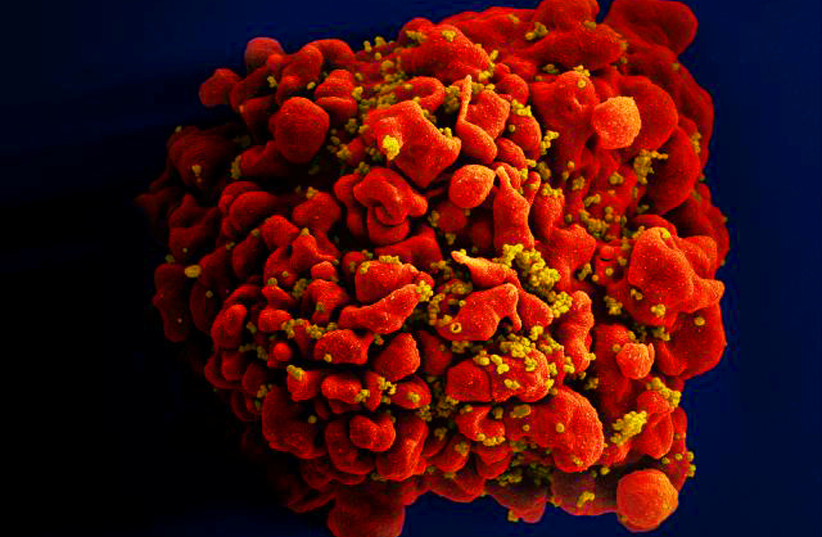
Scientists have pinpointed a hiding place for latent HIV in the human body in a new significant breakthrough in HIV research.
In a new study, the scientists confirmed that microglial cells (MG), a type of immune cell in the brain, can serve as a breeding ground for the virus.
Microglial cells are specialized immune cells that have a lifespan of approximately a decade in the brain. HIV can infect these cells, and some of them then transition into a dormant, latent state. If antiretroviral therapy (ART), the current treatment for HIV, is discontinued, the virus can rebound from these latent cells and reactivate the infection with the potential to revive the progression of HIV infection to AIDS.
By developing protocols to isolate brain microglia (MG) from humans on long-term, fully suppressive antiretroviral therapy (ART), researchers demonstrated the presence of replication-competent HIV, capable of progressing into AIDS, within these cells. This discovery has the potential to propel research that can eradicate HIV completely.
Isolating HIV's hideout in the brain
The peer-reviewed study employed intricate laboratory techniques in which the team overcame ethical obstacles regarding human brain tissue sampling and established protocols to isolate brain microglia (MG) and characterize viral reservoirs, or breeding grounds, in the non-human primate (NHP) model of infection. The team first studied the brains of macaques with the simian immunodeficiency virus (SIV), which is closely related to HIV. They developed techniques to isolate and separate pure brain myeloid cells from CD4+ cells, the key coordinators in immune response when HIV attacks, in the brain tissue.
Applying these techniques, researchers used samples from the "Last Gift" study at the University of California San Diego (UCSD) which recruits people with HIV (PWH) with terminal illnesses who wish to contribute to HIV cure research through tissue donation for rapid research autopsies within six hours of death. The team achieved successful isolation and ex vivo culturing of MG, setting the stage for a deeper understanding of HIV's persistence in the CNS.
"This had been suspected in the past, but proof in humans was lacking," said Yuyang Tang, PhD, the study's first author. "Our method for isolating viable brain cells provides a new framework for future studies on reservoirs of the central nervous system, and, ultimately, efforts towards the eradication of HIV.”
A key objective of the study served to confirm that the replication-competent HIV was truly derived from the brain MG. To achieve this, the researchers utilized a highly sensitive reverse transcription-quantitative polymerase chain reaction (RT-qPCR) and droplet digital polymerase chain reaction (ddPCR) assays, showing undetectable RNA for the T cell marker CD3 in isolated MG, thus demonstrating the purity of the sample. In stark contrast to the negative results in CNS T cells, replication-competent HIV was recovered from the isolated MG, representing the first direct evidence of its kind.
HIV latency in brain microglia
The study found that proviral HIV in the brain MG is largely silent in PWH on ART, posing a significant challenge to HIV eradication efforts. With the knowledge that latent HIV can hide in brain microglia, researchers are now investigating ways to target this reservoir. They suspect that HIV in the brain has adapted unique characteristics to replicate and hide from immune clearance.
"HIV is very smart," said Guochun Jiang, PhD, the study's senior author. "Over time, it has evolved to have epigenetic control of its expression, silencing the virus to hide in the brain from immune clearance. We are starting to unravel the unique mechanism that allows latency of HIV in brain microglia."
The researchers did discover that HIV latency in the brain MG could be disrupted by epigenetic modulators, highlighting the potential of these agents for targeted therapeutic interventions.
Implications and future directions
The research emphasizes the role of brain MG as a stable reservoir of persistent HIV, suggesting that future HIV eradication efforts need to address the possible persistence of the virus within CNS MG. Despite being limited by the small number of human samples available, these groundbreaking findings align with nonhuman primate studies and offer significant contributions to the scientific understanding of HIV's ability to persist within the CNS.
In their future work, the researchers aim to understand why latent HIV in the brain is not affected by NF-κB signaling, a key pathway controlling HIV expression elsewhere in the body. They are also working to determine the true size of the latent HIV brain reservoir.
"You want to be able to get it all, so it won't come back," said David Margolis, MD, co-author of the study and director of the UNC HIV Cure Center.